Research in the Terhune lab is focused in several complimentary areas:
Understanding the evolution and function of primate, modern human, and fossil hominin skull shape
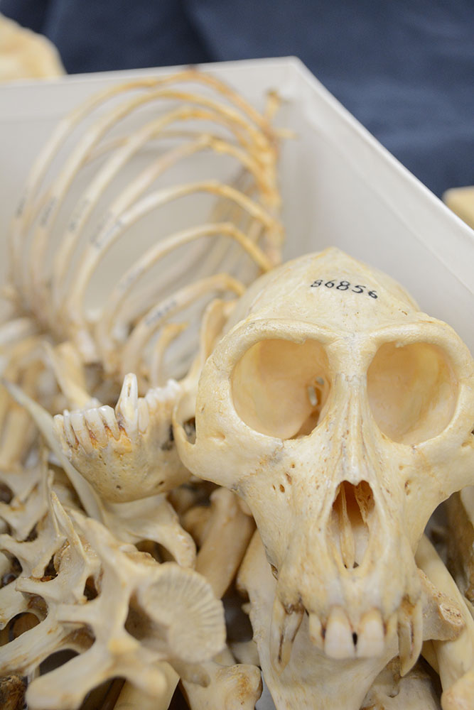
Primates range tremendously in size, from the mouse lemur to the gorilla. Primates also occupy a variety of niches across the globe, from tropical forests to desert savannahs, and in the case of humans, everywhere in between. Given this variability in size and geographical distribution, it then comes as no surprise that primates are also highly variable in their diet and feeding behaviors, with some species focusing very narrowly on a particular food resource, while other species are able to adapt their diet across a range of environments and food sources. This extreme variability in size, behavior, and diet has important implications for what the soft and hard tissues of these primates look like, and how species vary. In the Terhune lab, we focus primarily on evaluating shape variation in the primate, human, and fossil hominin skull. This research can be further broken down into three main areas:
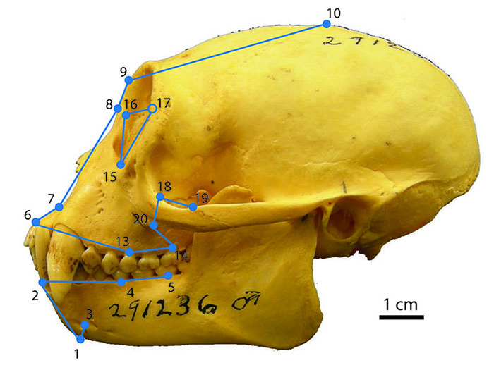
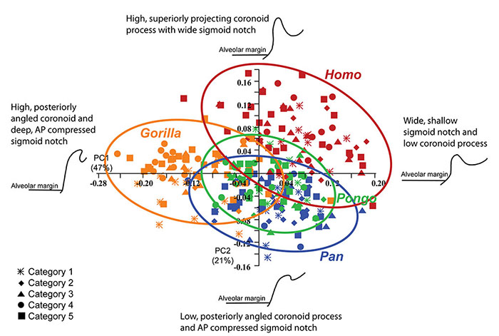
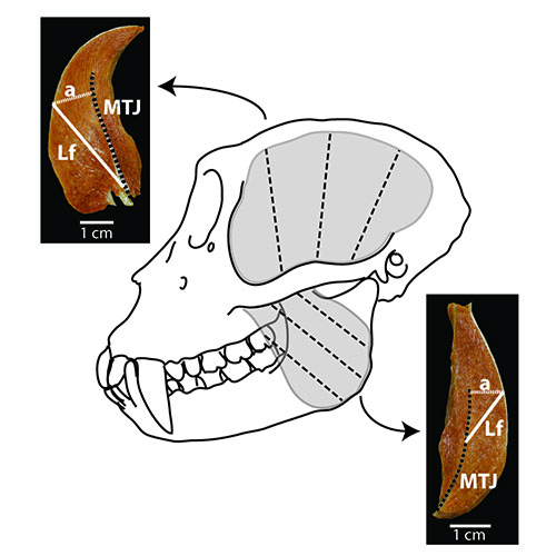
- Examining how primate skull shapes are linked to the function of the chewing apparatus and particularly the temporomandibular joint (TMJ).
- Investigating how the unique form of the modern human skull and chewing apparatus evolved during the past ~7 million years through comparative analyses of fossil hominins.
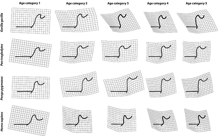
- Evaluating how skull shape changes during growth and development, and understanding what these shape changes might mean for hypotheses regarding taxonomy in fossil hominins, and for understanding variation in skull shape across living primates.
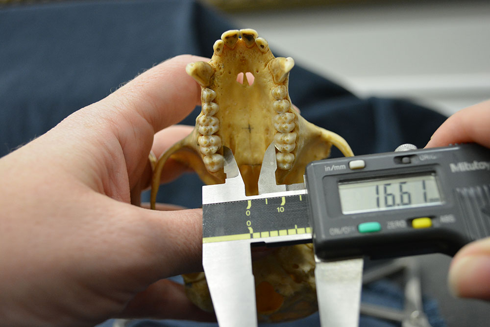
New and ongoing research projects are focused on studies of covariation between the TMJ and the dentition; linking TMJ and masticatory form to kinematic data describing primate mandibular movements during chewing; examining the internal architecture of the glenoid fossa and mandibular condyle to gain insight into loading of the TMJ; and examining the pathophysiology of TMJ remodeling and degeneration and the relationship to dental wear and oral trauma.


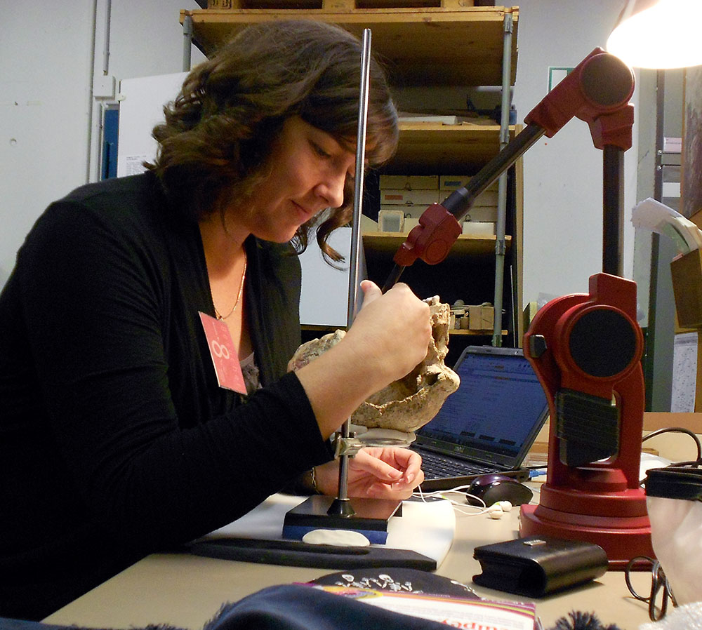
Advancing quantitative techniques for capturing the vast array of skull shapes of living primates.
The skull is a complex, three-dimensional (3D) structure, and contains a wealth of information regarding function and phylogenetic history. Teasing apart these different influences over skull shape is a complicated endeavor. Advanced quantitative techniques can help to capture this complex shape variation, and these methods (particularly those of the geometric morphometric toolkit) form a major component of the work being undertaken in the lab. Equipment available in the lab includes a microscribe digitizer, surface scanner, and advanced software for processing and analyzing CT and microCT data, as well as surface scans and 3D landmark data.


Evaluating models of hominin migration(s) into Europe and Asia during the early Pleistocene.
 The early Pleistocene is a critical time frame for understanding human evolution, and particularly for evaluating models of early human dispersal out of Africa and into Europe and Asia. Though the earliest appearance of hominins in Eurasia is dated to ~1.85 Ma, the best-dated hominins in Europe are not found until closer to 1.4 Ma. Given this relatively late arrival of human ancestors in Europe, it might be hypothesized that either there were geographic or environmental barriers that prevented hominins from dispersing into Europe at this time, or perhaps we simply haven’t yet identified sites with convincing evidence of an earlier hominin presence in Europe. One region that remains relatively less intensively explored is that of Eastern Europe, and few well documented faunal datasets describing environmental conditions in Eastern Europe during the early Pleistocene are available. Research in the lab is therefore also focused on evaluating models of hominin migration (or perhaps multiple migrations) into and out of Europe during the early Pleistocene, particularly from the perspective of Eastern Europe.
More information about our ongoing field project, a Pleistocene survey of the Olteţ River Valley, Romania, can be found on the Olteţ River Valley research page.
A lot of the current research in the Terhune Lab utilizes micro-computed tomography (microCT) imaging. MicroCT uses x-rays to non-destructively generate high-resolution two- and three-dimensional (2D/3D) representations of an object’s internal and external structure, providing exquisite details of the internal structure of the object being scanned. We are particularly interested in using microCT and diceCT (diffusible-iodine contrast enhanced CT) to examine the internal structure of bony and soft-tissue anatomy of the primate masticatory apparatus and skull. This work is primarily being conducted at the University of Arkansas MicroCT Imaging Consortium for Research and Outreach (MICRO), which houses a Nikon X TH 225 ST microCT imaging system. You can learn more about this facility by visiting the MICRO webpage.